PELVIC/ACETABULAR SURGERIES
Overview
Pelvic fractures and acetabular fractures are among the most serious injuries treated by orthopedic surgeons. Often the result of a traumatic incident such as a motor vehicle accident or a bad fall, these fractures require rapid and precise treatment and, in some cases, one or more surgical procedures. People of all ages are vulnerable to these injuries. In addition, some elderly patients with fragile bones due to osteoporosis develop pelvic fractures and fractures of the acetabulum with a lower impact fall.
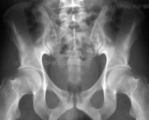
Radiograph of a normal pelvis
Radiograph of the pelvis demonstrating a fracture of the pubic bone
The complex nature of these fractures can be better understood by looking at the anatomy that is involved. The pelvis is made up of several bones (ileum, ischium and pubic bones) which create a bony ring, meeting at the pubic symphysis in the front and the sacrum (a bone situated at the lower end of the spine) in the back. Together with a number of ligaments and muscles, the bones of the pelvis support the weight of the upper body and rest on the hip joints. The pelvis protects abdominal organs including the intestines and the bladder, as well as major nerves and blood vessels. Pelvic fractures may occur at any location on the bones depending on the nature of the accident and the areas of impact.
The
Acetabulum refers to the part of the pelvis that meets the upper end of the thigh bone (the femoral head) to form the hip joint. In a healthy hip, these two bones fit together like a ball and cup, in which the ball rotates freely in the cup. Cartilage lines the bones where they meet at the joint and there is little friction between the surfaces during movement.

Anatomical illustration of the acetabulum
The term broken hip usually refers to a fracture of the ball portion of this joint, that is, the upper femur, femoral neck or the femoral head. In this section, we are speaking specifically of a fracture of the cup or acetabulum. Fractures of the acetabulum are harder to treat because access to this bone is more difficult, and because of the acetabulum's proximity to the major blood vessels to the legs, the sciatic nerve (the major nerve that arises from the lower spine and provides sensation and movement to the leg and foot), the intestines, the ureter and the bladder. Unlike a hip fracture, which can be treated relatively easily, to repair an acetabular fracture, the orthopaedic surgeon, must, in essence, fix the broken bones from the inside out.
In fractures of this type, the femoral head is often driven through the acetabulum because of the impact of the fall or accident. If the femoral head ends up outside the acetabulum, this is known as a dislocation of the hip joint. Some patients have both a fracture and a dislocation.

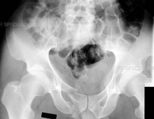
Anatomical illustration of the acetabulumRadiograph of the left hip demonstrating a posterior dislocation of the hip with an associated
Posterior Wall type fracture of the acetabulum
Unfortunately, patients with fractures of the pelvis and/or acetabulum, almost always also experience serious injury to surrounding soft tissue (skin and muscles) and neurovascular structures (nerves, arteries and veins). In addition, especially in the case of pelvic fractures, adjacent organs can be seriously injured. With both types of fracture, there is significant bleeding and risk of nerve damage.
In patients with multiple injuries, treatment begins with the trauma team at the scene, and then subsequently in the emergency room--a team of general surgeons, anesthesiologists and nurses--who work together to control bleeding, address damage to the head and chest, and other organs that may have been affected, such as the bladder and intestines, and to stabilize broken bones. During this early resuscitation phase of treatment, the orthopaedic surgeon may need to stabilize the fracture by using an external frame to temporarily hold the bones in proper alignment while other problems are treated. This is called temporary external fixation. Surgeons construct these frames using steel pins that are inserted into the bone and joined together by clamps and rods and can do so very rapidly.

ARadiograph of the pelvis demonstrating application of a pelvic external fixator.
Once the patient is stabilized--bleeding has stopped and other life-threatening injuries have been addressed--the fractures can be treated definitively. Successful treatment for both of these types of fractures requires the skills of an interdisciplinary team, with orthopaedic surgeons working closely with the trauma team (general surgeons), the anesthesiologists and nurses. Following surgery, rehabilitation specialists play a key role in recovery.
Because of the complex nature of these fractures and because many orthopedic surgeons do not regularly treat them, patients who initially go to a community hospital for emergency attention are often transferred to an institution that specializes in such injuries.
Surgical Treatment
Realignment of the bones may be done either as an open reduction, in which the orthopaedic surgeon makes an incision to directly manipulate the bone, or as a closed reduction, in which this incision is not necessary. Once the bones are realigned, the surgeon uses internal or external fixation to hold the bone in proper position during healing. Metallic devices including wires, pins, screws, and plates are used.

Radiograph of the pelvis following open reduction and internal fixation (ORIF) of a complex comminuted fracture of the left acetabulum, hemipelvis and pubic symphysis.
Patients with pelvic fractures may require one or more surgical procedures. The surgeon may begin with an External Fixation (Ex-Fix) technique in which an open or closed reduction is performed and the bones are then held in place using an external fixator, or frame. This is done by threading pins into the bone on either side of the fracture. These pins are then connected to rods outside the skin, which form a frame.
While the Ex-Fix technique is sometimes the only procedure needed to repair a fractured pelvis, some patients require additional surgery or surgeries in which plates and screws are used internally to hold the bones in place. Depending on the site and complexity of the fracture, the surgeon may have to fix the front of the pelvis, the back of the pelvis, or both. Separate operations may be needed for each area that needs treatment.
Patients with acetabular fractures often require an Open Reduction with Internal Fixation (ORIF), especially those patients who also have displacement of the joint. The surgeon realigns or reduces the bones as precisely as possible to prevent the development of post-injury related problems, especially arthritis. The bones are rigidly fixed with plates and screws to prevent future displacement and allow for rehabilitation to begin as quickly as possible.
Fractures of the acetabulum are usually not treated for 5-10 days following the injury. Because the patient experiences significant bleeding with this fracture, the orthopaedic surgeon must wait for the patient's own clotting mechanisms to go into effect--usually within 3-5 days. During this period the patient may be in traction to prevent additional injury.
Pre-Operative Procedures
Patients scheduled for surgery undergo a number of tests. These include:
- Blood tests
- An electrocardiogram (or EKG) that tests the electrical activity of the heart
- A chest x-ray to ensure that the lungs have not been injured and have no fluid in them and that the patient has no infection of the lung, i.e. pneumonia
- Conventional radiographs, Computerized Tomography (CT scan), or Magnetic Resonance Imaging (MRI): Each of these tests helps the surgeon get as much information as possible about the fracture before beginning surgery. CT scans are particularly useful since they allow the physician to see the fracture in several planes and also see a 3-D model of the fracture on a computer monitor
- Magnetic Resonance Venogram (MRV): Assesses the patient's veins. Many patients with fractures of the pelvis and acetabulum develop blood clots in the veins of the pelvis, thighs or lower legs. If the clot travels through the body to the lungs it is called a pulmonary embolism and can interfere with the patient's breathing. If the MRV shows that a clot is present, treatment for the clot is immediately started. This may include placement of an Inferior Vena Cava Filter, i.e. a "strainer" in the major vein to the heart to prevent any blood clots going to the lungs (pulmonary embolism).
Post-Operative Care
Following surgery, managing the patient's pain and managing any complications that arise due to the injury are primary concerns.
Initially, pain medication will be given by injection. However, many patients are able to use a pump that controls the amount of pain medication given. This is known as Patient Controlled Analgesia (PCA) and offers patients the benefits of managing his or her pain. Since there is a maximum dose that can be delivered at any given time, there is no danger that the patient will receive too much medication.
Other medications that may be given include anticoagulants to thin the blood and avoid the development of blood clots, and Indocin, which prevents bone formation in areas around the muscles.
Patients are encouraged to get up and out of bed as soon as possible, since doing so helps to avoid some of the complications associated with these injuries. A regimen of physical therapy is followed to maintain muscle strength and range of motion during recovery.
After surgery to repair a pelvic fracture or fracture of the acetabulum, many patients continue to feel the effects of damage to nerves that might have occurred during the traumatic event or the surgery. Important branches of the lumbar and sacral nerves may be either stretched or torn, especially in the case of unstable pelvic fractures. Injuries to the nerves result in decreased feeling in a limb and/or difficulty or inability in moving part of the limb. It is difficult to predict whether these nerves will fully recover. However, the majority of patients do regain some sensation and function of the limb within six to eighteen months after their injury.
SPORTS MEDICINE INFORMATION
Sports injuries are injuries that occur to athletes in major sporting events. In many cases, these types of injuries are due to overuse of a part of the body when participating in a certain activity. For example, runner's knee is a painful condition generally associated with running, while tennis elbow is a form of repetitive stress injury at the elbow, although it does not often occur with tennis players. Other types of injuries can be caused by a hard contact with something. This can often cause a broken bone or torn ligament or tendon

Injuries are a common occurrence in professional sports and most teams have a staff of Athletic Trainers and close connections to the medical community. Controversy has arisen at times when teams have made decisions that could threaten a players long-term health for short term gain.
Classification
Sports injuries can be broadly classified as either traumatic or overuse injuries. Traumatic injuries account for most injuries in contact sports such as Football, Rugby, Australian rules football, Gaelic football and American football because of the dynamic and high collision nature of these sports. These injuries range from bruises and muscle strains, to fractures and head injuries.
A bruise or contusion is damage to small blood vessels which causes bleeding within the tissues. A muscle strain is a small tear of muscle fibers and a ligament sprain is a small tear of ligament tissue. The body’s response to these sports injuries is the same in the initial five day period immediately following the traumatic incident - inflammation.
Inflammation is characterized by pain, localized swelling, heat, redness and a loss of function.
